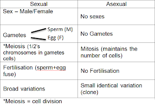
Examples of mitosis:
http://highered.mcgraw-hill.com/sites/0072495855/student_view0/chapter2/animation__mitosis_and_cytokinesis.html
http://cellsalive.com/mitosis.htm
Video of mitosis
Another mitosis video in 3D
Monday, 29 August 2011
3.24 - Mitosis
a) Outline/summary
number or chromosomes in neucleus = diploid number (2n)
in humans 2n=46
in cats 2n=38
In the process of mitosis a cell divides into 2 identical cells:
-they have the same number of chromosomes
-same set of chromosomes
Each new cell has a diploid nucleus
b) Details
Copying the chromosomes is a process called DNA replication
-Each chromosome undergoes a copying process to form an identical copy of itself (same genes)
-These 2 copies are held together by the structure around the centre region, know as the centromere
-These are known as a pair of chromatids
-The process (DNA replication) takes place in the nucleus (the process cant be seen because the nucleus stays intact). this is know as the interphase
c) Stages of mitosis
We would normally see that the nucleus has a spherical structure if we looked at it down a microscope and we would be unable to see the chromosomes.
It is during that interface that DNA replication occurs
Prophase- The membrane breaks down and the chromosomes (pair of chromatids) can be seen. each chromosome got copied
The nucleus is gone and there is a network of protein molecules inside the cell called a spindle
-These go from one pole of a cell to the other
-The pair of chromatids will join on one of the spindles fibres at the centromere
Metaphase: When the pair of chromatids are attached to a spindle fibre by the centrometer
-The chromatid is in the middle across the equator of the cell
Anaphase: the spindle fibre shortens pulling one chromatid in one direction and the other chromatid in the other direction. This pulls the chromatides to the poles of the cell.
-This seperated the chromatids
Telophase: Nucleus begins to reform around the chromosomes at either end of the cells (new nucleus of the new cell)
We see the formation of 2 nuclei at opposite ends of the cell
Cytokinesis: The cell splits into 2 (not a part of mitosis)
Thursday, 25 August 2011
3.16 - DNA, nature of genetic code
DNA is shaped as a double helix, they appear to be parrallel
In the center there are bases:
-Adenine (A)
-Thymine (T)
-Cytosine (C)
-Guanine (G)
These molecules hold the two helices together, they are held together by bonding:
A - T
C - G
The base pairs hold one side of the helix to the other
the order of the bases (the gene): ACTGAACCAG
3.15 - Genes
Genes: A section of a molecule of DNA
The gene is the characteristic of an organism
Genes are located in the nucleus
The information is then passed to the cytoplasm, the genetic information is transformed into a protein.
3.14 - Chromosomes
Chromosomes: genetic information within a cell
Chromosmes is composed of a molecule called DNA
DNA has a shape known as the double helix
section's of DNA are called genes, a chromosome is composed of 1000's of genes (DNA = 3.15)
each gene has the information for the construction of the protein (see 3.16)
The protein gives the characteristics associated with the gene (eg. Blood group)
Different organisms have different numbers of chromosomes (eg cat = 38, chicken = 78, chimp = 42, humans=46 chromosomes per cell)
Chromosomes are known to work in pairs (homologous pairs)
(3.17)
Saturday, 20 August 2011
Subscribe to:
Comments (Atom)


